ปัจจุบันเรียกได้ว่ากระแสของปัญญาประดิษฐ์ Artificial intelligence (AI) ถือเป็นเทคโนโลยีที่ผู้คนทั่วโลกต่างให้ความสนใจและจับตามอง จนทุกวันนี้ AI ได้เข้ามามีส่วนสำคัญในการวิเคราะห์ข้อมูลที่มีอยู่จำนวนมหาศาล เพื่อช่วยพัฒนาธุรกิจและอุตสาหกรรมประเภทต่าง ๆ และเกี่ยวข้องกับชีวิตมนุษย์ในหลากหลายด้านมากขึ้น หนึ่งในนั้นคือด้านการแพทย์ ซึ่งปฏิเสธไม่ได้ว่า AI ถือเป็นกลไกสำคัญในการยกระดับทางการแพทย์ให้เกิดประสิทธิภาพมากยิ่งขึ้น
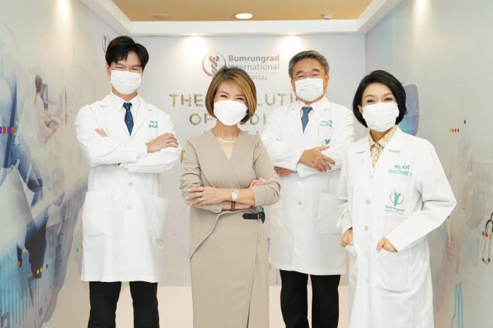
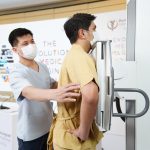
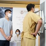
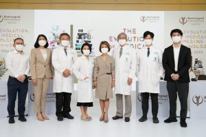
โดยในงานแถลงข่าว The Evolution of Medical Imaging Technology เภสัชกรหญิงอาทิรัตน์ จารุกิจพิพัฒน์ ประธานเจ้าหน้าที่บริหาร โรงพยาบาลบำรุงราษฎร์ ได้กล่าวถึงวิสัยทัศน์และทิศทางของบำรุงราษฎร์ ที่มุ่งให้ความสำคัญด้านนวัตกรรมทางการแพทย์มาอย่างต่อเนื่อง จะเห็นได้ว่าที่ผ่านมา บำรุงราษฎร์ได้เปิดรับเอานวัตกรรมเทคโนโลยีใหม่ๆ ที่เหมาะสมเข้ามาใช้ในโรงพยาบาลเป็นอันดับต้นๆ ของประเทศ ทั้งในส่วนของการศึกษาวิจัย และการนำนวัตกรรมและเทคโนโลยีทางการแพทย์ขั้นสูงเข้ามาใช้ โดยเฉพาะเรื่อง AI, Big Data, การศึกษาเกี่ยวกับยีน (Genomics) และ การส่งเสริมสุขภาพเชิงวิทยาศาสตร์ (Scientific Wellness) เพื่อเอื้อประโยชน์ต่อทั้งแพทย์ผู้ให้การรักษา บุคลากรทางการแพทย์ สหสาขาวิชาชีพ ในการบริบาลผู้ป่วยอย่างครอบคลุมทุกมิติในทุกกระบวนการของการรักษาเพื่อให้เกิดผลลัพธ์ที่ดีที่สุด โดยคำนึงถึงความปลอดภัยของผู้ป่วยเป็นสำคัญ
สำหรับการนำ AI มาใช้ในด้านรังสีวิทยาที่บำรุงราษฎร์นั้น ถือว่ามีความโดดเด่นมาก เนื่องจากมีการนำเทคโนโลยีมาใช้เป็นแผนกแรก ๆ ในโรงพยาบาล เรียกได้ว่า ‘รังสีแพทย์’ เป็นแถวหน้าด้านการแพทย์ในยุคดิจิทัล ซึ่งหากมองย้อนกลับไปตั้งแต่ปี 2543 หรือ 22 ปีก่อน บำรุงราษฎร์ได้เริ่มใช้งานระบบ Picture Archiving and Communication System (PACS) ซึ่งเป็นระบบที่ใช้ในการจัดเก็บรูปภาพทางการแพทย์ (Medical Images) หรือภาพถ่ายทางรังสี โดยมีการรับส่งข้อมูลภาพในรูปแบบดิจิทัลเป็นครั้งแรก ต่อมาในปี 2544 นับเป็นปีแรกที่เริ่มนำเทคโนโลยีการถ่ายภาพแบบ Computed Radiography คือการใช้แผ่นรับภาพหรือเรียกว่า image plate แทนการใช้ฟิล์ม โดยมีเครื่องอ่านภาพ (image reader) เพื่ออ่านข้อมูลบนแผ่นก่อนนำส่งผ่านระบบคอมพิวเตอร์ โดยแพทย์สามารถอ่านผลได้ทั้งการสั่งพิมพ์ฟิล์มหรือบนคอมพิวเตอร์และต่อมาในปี 2550 ได้พัฒนาเป็นดิจิตัลเต็มรูปแบบ โดยใช้ระบบเซ็นเซอร์หรือตัวรับภาพขนาดใหญ่ (detector) แทนการใช้แผ่นรับภาพแบบเดิม โดยประมวลผลและทราบผลภาพในเวลาเพียงเสี้ยววินาทีโดยไม่ต้องมีอุปกรณ์อ่านข้อมูล
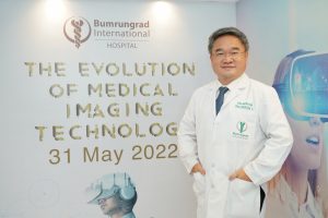
นพ. อลงกรณ์ เกียรติดิลกรัฐ รองหัวหน้าแผนกรังสีวิทยา และแพทย์ชำนาญการด้านรังสีวิทยา โรงพยาบาลบำรุงราษฎร์ เปิดเผยว่า “ล่าสุด บำรุงราษฎร์ได้มีการนำ Radiology AI ปัญญาประดิษฐ์ทางรังสีวิทยาเข้ามาใช้ในโรงพยาบาล เสมือนเป็นผู้ช่วยรังสีแพทย์ทั้งในส่วนของการคัดกรอง การวิเคราะห์ และการวินิจฉัย พร้อมระบุตำแหน่งภาวะความผิดปกติของปอด และมะเร็งเต้านมระยะแรกเริ่ม โดย Radiology AI ถือเป็น Deep Learning AI ซึ่งเป็นโมเดลการเรียนรู้ในเชิงลึก เป็นอัลกอรึทึมที่ใช้โครงข่ายใยประสาทเสมือน ด้วยการเลียนแบบการทำงานระบบประสาทของมนุษย์ที่มีเซลล์ประสาทเชื่อมต่อกัน ฉะนั้น Radiology AI จะสื่อสารโดยการประมวลผลที่ละเอียดลึกซึ้งกว่า Machine Learning รวมถึงสามารถประมวลผลต่อได้เอง ซึ่งเทคโนโลยีนี้ได้รับการรับรองจากองค์การอาหารและยาแห่งสหรัฐอเมริกาในปี 2564 ที่สำคัญ Radiology AI มีความล้ำทันสมัยในการวินิจฉัย มีความเสถียร แม่นยำ และรวดเร็ว ด้วยการใช้ Microsoft Azure แพลตฟอร์มคลาวด์คอมพิวติ้ง ซึ่งเป็นที่ยอมรับในระดับโลก
ทั้งนี้ บำรุงราษฎร์ได้ใช้เทคโนโลยี Radiology AI ในศูนย์ตรวจสุขภาพ แผนกผู้ป่วยวิกฤต (ICU) แผนกฉุกเฉิน และแผนกอื่น ๆ โดยแบ่งเป็น 2 ส่วน คือ 1. Radiology INSIGHT CXR จะใช้ในการวิเคราะห์ภาพเอ็กซ์เรย์ทรวงอก เฉลี่ย 100,000 ภาพต่อปี และ 2. Radiology INSIGHT MMG จะใช้วิเคราะห์มะเร็งเต้านมจากภาพถ่ายแมมโมแกรม โดยบำรุงราษฎร์มุ่งหวังที่จะส่งมอบการบริการแก่ผู้ป่วยอย่างมีคุณค่าที่สุด โดยการตรวจหามะเร็งในเวลาที่เหมาะสมเพื่อความสำเร็จในการรักษา ซึ่งนอกจากความชำนาญการและประสบการณ์ของรังสีแพทย์แล้ว Radiology AI ยังมีส่วนช่วยสนับสนุนให้บำรุงราษฎร์ขับเคลื่อนนวัตกรรมทางการแพทย์ที่ก้าวล้ำสู่ระดับโลกอีกด้วย

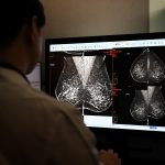
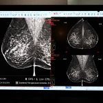
พญ. พัชรี ประสิทธิ์วรนันท์ แพทย์ชำนาญการด้านรังสีวิทยา โรงพยาบาลบำรุงราษฎร์ กล่าวว่า “จากข้อมูลสถาบันมะเร็งแห่งชาติ ในปี 2563 พบหญิงไทยป่วยด้วยมะเร็งเต้านมรายใหม่ ประมาณ 18,000 คนต่อปี และมีผู้เสียชีวิตจากมะเร็งเต้านม ประมาณ 4,800 คน หรือเฉลี่ย 13 คนต่อวัน ซึ่งมะเร็งเต้านมเป็นอันดับหนึ่งของมะเร็งที่พบบ่อยในผู้หญิงไทยและจากรายงานยังมีแนวโน้มสูงขึ้นทุกปี ฉะนั้นผู้หญิงในวัย 40 ปีขึ้นไป จึงควรเข้ารับการตรวจแมมโมแกรมเป็นประจำทุกปี ปัจจุบันบำรุงราษฎร์มีการนำปัญญาประดิษฐ์สำหรับแมมโมแกรม หรือ Radiology INSIGHT MMG ซึ่งมีส่วนช่วยในการคัดกรอง วิเคราะห์ และวินิจฉัยมะเร็งเต้านมบนภาพถ่ายแมมโมแกรม โดยก่อนหน้านี้ รังสีแพทย์ก็มีการอ่าน แปลผล วิเคราะห์ และวินิจฉัยโรคจากภาพถ่ายต่างๆ อยู่แล้ว แต่การนำ AI เข้ามาใช้ทางการแพทย์นั้น ก็เสมือนมีผู้ช่วย มาช่วยวิเคราะห์ โดยในส่วนนี้เองจะช่วยเพิ่มความรอบคอบให้กับแพทย์ได้มากขึ้น ทั้งนี้ รังสีแพทย์จะนำผลของ Radiology AI มาประกอบการวินิจฉัย มะเร็งเต้านมหากได้รับการวินิจฉัยตั้งแต่ระยะแรกเริ่ม และเริ่มการรักษาอย่างเหมาะสมทันท่วงที ก็จะทำให้ผู้ป่วยมีอัตราการรอดชีวิต 5 ปี สูงถึง 99% แต่หากวินิจฉัยช้า ทำให้อัตราการรอดชีวิต 5 ปี จะลดลงเหลือ 86% ลงมาจนถึง 29% ตามระยะของมะเร็งที่สูงขึ้น ดังนั้น หากตรวจพบเร็วจะช่วยเพิ่มโอกาสความสำเร็จในการรักษาผู้ป่วยได้มากขึ้น”
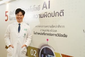
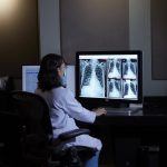
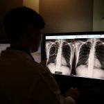
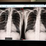
ด้าน นพ. กุรุวินท์ ลิ้มสมุทรเพชร แพทย์ชำนาญการด้านรังสีวิทยา โรงพยาบาลบำรุงราษฎร์ กล่าวถึงปัญญาประดิษฐ์สำหรับการเอกซเรย์ปอด หรือ Radiology INSIGHT CXR เสมือนเป็นผู้ช่วยรังสีแพทย์ในการตรวจหาภาวะความผิดปกติของภาพเอกซเรย์ปอด เช่น ก้อนมะเร็งปอด ซึ่งรวมถึงจุดเล็กๆ ในตำแหน่งที่ยากต่อการวินิจฉัย วัณโรคในระยะที่แสดงอาการ รวมถึงช่วยวินิจฉัยภาวะฉุกเฉินได้ตั้งแต่เนิ่น ๆ เช่น ภาวะลมรั่วในปอด ซึ่งหากเกิดลมรั่วน้อยๆ ผู้ป่วยบางรายอาจยังไม่แสดงอาการใด ๆ หรืออาจมาด้วยอาการเจ็บหน้าอก แต่หากรังสีแพทย์สามารถวินิจฉัยได้เร็วและทราบผลตั้งแต่เนิ่น ๆ ก็จะทำให้ผู้ป่วยไม่พลาดโอกาสในการรักษา โดยมีงานวิจัยพบว่าผู้ป่วยโรคมะเร็งปอด หากได้รับการวินิจฉัยตั้งแต่ระยะแรกเริ่ม จะทำให้มีอัตราการรอดชีวิต 5 ปี ถึง 73% แต่หากวินิจฉัยช้า อัตราการรอดชีวิต 5 ปีจะเหลือเพียง 18% เท่านั้นอย่างไรก็ตาม Radiology AI ไม่ได้เข้ามาเพื่อทดแทนที่รังสีแพทย์ แต่จะเป็นเครื่องมือสนับสนุนสำหรับกระบวนการวินิจฉัยและตัดสินใจของรังสีแพทย์ โดยจะเป็นรูปแบบการให้ “ความเห็นที่สอง” (Second Opinion) ซึ่งจากผลการสำรวจความคิดเห็นของรังสีแพทย์ส่วนใหญ่มากกว่า 60% ระบุว่าได้รับประโยชน์จากการนำ Radiology AI มาใช้ในการช่วยวิเคราะห์ภาพเอกซเรย์ปอด สำหรับในส่วนของซอฟต์แวร์ Radiology AI นั้น ยังมีโอกาสการพัฒนาอย่างต่อเนื่องเพื่อให้ตอบโจทย์การทำงานที่มีประสิทธิภาพมากขึ้น ขณะเดียวกัน ความคิดเห็นและข้อเสนอแนะของรังสีแพทย์ของโรงพยาบาลบำรุงราษฎร์ มีความสำคัญและมีส่วนช่วยในการปรับปรุงประสิทธิภาพการทำงานของ Radiology AI ให้ดียิ่งขึ้นแก่บริษัทผู้พัฒนาปัญญาประดิษฐ์ นอกจากนี้ Radiology AI นี้ ยังช่วยเสริมให้การทำงานร่วมกันระหว่างรังสีแพทย์, AI และแพทย์ผู้รักษาผู้ป่วย มีประสิทธิภาพและเกิดประสิทธิผลในการรักษาสูงสุดต่อผู้ป่วย อีกทั้งยังเป็นตัวเร่งการเรียนรู้ของรังสีแพทย์รุ่นใหม่ ๆ ในเวลาจำกัดได้อย่างมีประสิทธิภาพ
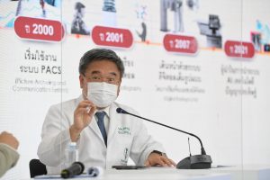
นพ. อลงกรณ์ เกียรติดิลกรัฐ กล่าวปิดท้ายถึงความร่วมมือในอนาคต บำรุงราษฎร์มีแนวโน้มที่จะร่วมมือกับบริษัทผู้พัฒนา Radiology AI ในประเทศเกาหลีใต้ และโรงพยาบาลชั้นนำ 20 แห่งทั่วโลก เพื่อร่วมกันพัฒนา Radiology AI เพื่อให้มีความฉลาดมากยิ่งขึ้น ตรวจพบโรคได้เร็วขึ้น มีความแม่นยำมากขึ้น และสามารถขยายขอบข่ายการทำงานในการตรวจวินิจฉัยโรคให้ครอบคลุมมากขึ้น โดยแนวทางความร่วมมือนี้ นับเป็นจุดเริ่มต้นที่สำคัญอย่างมากต่อระบบการแพทย์ยุคใหม่ ซึ่ง AI ถือเป็นตัวแปรสำคัญที่จะยกระดับความสำเร็จให้กับวงการแพทย์ในทุกภาคส่วน อีกทั้งยังช่วยส่งเสริมการมีคุณภาพชีวิตที่ดีของผู้คนทั่วโลกอีกด้วย
Bumrungrad launches Radiology AI–assisting radiologists in analyzing & locating abnormalities in the lungs and detecting breast cancer.
At present, Artificial intelligence (AI) has attracted global interest because it can analyze large amounts of data to revolutionize businesses and industries, especially in the medical sciences. Further, it is indisputable AI plays a key role in increasing medical competency.
In the press conference “The Evolution of Medical Imaging Technology”, Pharmacist Artirat Charukitpipat, CEO of Bumrungrad International Hospital, discussed Bumrungrad’s vision and direction to continue creating innovative clinical experiences. Bumrungrad is one of the top hospitals in Thailand in terms of the adoption of the latest medical technology. We do research and use advanced medical technology including AI, Big Data, Genomics, and Scientific Wellness to assist our physicians and multidisciplinary healthcare professionals to deliver holistic healthcare services, resulting in the best possible treatments which meet international standards of safety.
The Radiology Department of Bumrungrad is a standout in adopting AI to treat patients because it was the first department using the AI technology. Radiologists are frontline physicians in the digital age. However, 22 years ago in the year 2000, Bumrungrad began using the Picture Archiving and Communication System (PACS) to collect medical images – particularly radiographs, to share with our staff for the first time as digital files. In 2001, Bumrungrad adopted Computer Radiography, which uses digital image plates to replace X-ray film radiography. The scanner then reads out the latent image from the plates before sharing them online. Physicians can either print the files on film or examine them on the computer. In 2007, the Bumrungrad Department of Radiology moved to digital radiography using digital detector arrays (DDAs), which provide high quality digital images and can upload them to our online system in seconds. There is no need for the scanner anymore.
Dr. Alongkorn Kiatdilokrath, Assistant Chairperson of Department of Radiology and Radiologist at Bumrungrad International Hospital, reveals, “Recently, Bumrungrad has adopted Radiology AI, which assists radiologists in screening, analyzing, and locating abnormalities in the lungs and detecting early stages of breast cancer. The Radiology AI deep learning algorithm forms large artificial neural networks to imitate how the human brain and the nervous system work. Hence, Radiology AI offers more accurate imaging and interpretation than other forms of machine learning, which only performs tasks based on past data. Moreover, Radiology AI can perform further data analysis and was approved by the U.S. Food and Drug Administration in 2021. Radiology AI is stable, accurate, and fast because it uses Microsoft Azure, an internationally recognized cloud computing platform.
Currently, Bumrungrad is actively using the Radiology AI in the check-ups, intensive care unit, emergency room, and more. There are two uses of the Radiology AI. Firstly, Radiology INSIGHT CXR analyzes an average of 100,000 chest x-ray images per year. Secondly, Radiology INSIGHT MMG supports the analysis of mammographic images to detect breast cancer. Through AI, Bumrungrad aims to deliver a meaningful patient journey by timely detecting cancer. With the expertise of our radiologists, the Radiology AI will further support Bumrungrad to drive international groundbreaking medical innovation.
Dr. Patcharee Prasitvoranant, Radiologist at Bumrungrad International Hospital, states, “In 2020, the National Cancer Institute reported 18,000 new cases of breast cancer in Thai women. There were 4,800 deaths from breast cancer, or 13 deaths per day on average. Breast cancer is still the most common form of cancer affecting Thai women and the number of cases rises annually. Women over the age of 40 should have a yearly mammogram. Currently, Bumrungrad uses Radiology INSIGHT MMG to screen, analyze, and diagnose early-stage breast cancer through mammographic images. In the past, radiologists read, interpreted, analyzed, and diagnosed medical problems just through imagery. However, the Radiology AI in medical technology is an adjunctive tool helping radiologists to analyze the images, making diagnoses more accurate. Radiologists diagnose breast cancer using the results of Radiology AI. If breast cancer is diagnosed early and the treatment process can start immediately, the 5-year survival rate is as high as 99%. If it is diagnosed later, the 5-year survival rate can decrease to as little as 86% to 29%, depending on how far the cancer has progressed. In sum, early diagnosis means a higher chance of successful treatment.”
Dr. Kuruwin Limsamutpetch, Radiologist at Bumrungrad International Hospital, adds, “The Radiology INSIGHT CXR is like an assistant to radiologists in spotting abnormalities in the lung – like malignant tumors, including small ones hidden in spots that are hard to diagnose. Moreover, Radiology INSIGHT CXR can detect active tuberculosis and diagnose emergency cases like pneumothorax. At the onset of pneumothorax, when air leaks just a little bit into the space between the lung and chest wall, the patient may not have any symptoms or just mild chest pain. If radiologists can diagnose it early and the Radiology AI results come quickly, the patient will not miss the chance to be treated. Research findings show that early diagnosed, lung cancer patients have a 5-year survival rate of 73%. But if it is diagnosed later, the rate drops to just 18%.
However, Radiology AI is not meant to replace the radiologist. It is a tool which assists diagnosis and radiologist decisions. In other words, it gives a second opinion. A survey found that more than 60% of radiologists find the Radiology AI helpful in helping to analyze the lung images. The Radiology AI software is in active development to increase its efficacy. Radiologist feedback at Bumrungrad is crucial to improving Radiology AI’s competency. The Radiology AI encourages radiologists, AI, and other physicians who treat the same patients to work cohesively as a team, resulting in effective treatments. The Radiology AI also successfully increases the pace of learning for new radiologists.
Dr. Alongkorn Kiatdilokrath concludes that in the future, Bumrungrad intends to form a partnership with the Radiology AI developer in South Korea and 20 internationally recognized hospitals to increase the effectiveness of Radiology AI by enabling it to diagnose disease faster and more accurately. The goal is to enable radiologists to diagnose a wider range of conditions. The partnership marks a milestone in modern medical technology, in which AI is an important factor for successful holistic medicine. The AI also improves the wellbeing of people worldwide.


